Uniqueness
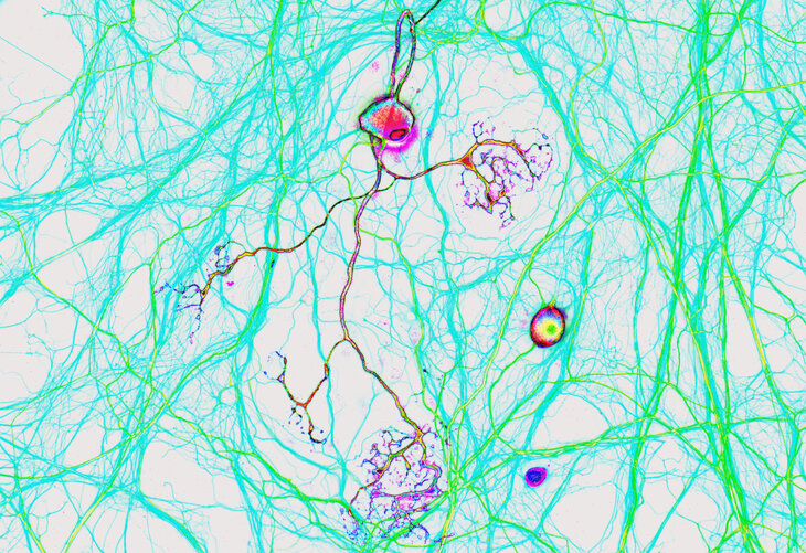
Photomicrograph of neurons stained with SYSY Antibodies: Parvalbumin (195 308), Neurofilament H (171 108) and beta3-Tubulin (302 311).
What makes you unique? Is it your color? Your shape? Or is it what you have inside?
This artwork is derived from an immunocytochemistry image. A parvalbumin-specific neuron from the peripheral nervous system can be seen in a rainbow of colors, as can the soma of a second neuron. Many other neurites from parvalbumin-negative neurons whose cell bodies are not displayed in the picture can also be observed in green (labeled with an antibody to Neurofilament H) and light blue (labeled with an antibody to beta3-tubulin).
For more scientific information about the highlighted proteins and the antibodies used in this image, click here: Parvalbumin, Neurofilament H, beta3-Tubulin 311.